ASU and Phoenix Children’s Hospital Use 3D Tech for Cardiac Print Lab
ASU engineers and Phoenix Children’s Hospital have come together to develop a realistic cardiac print lab – a library of hearts -using a combination of medical imaging and 3D technology.
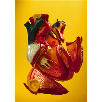
Led by associate professor of engineering at ASU, David Frake, the engineering team has developed an incredible system capable of creating 3D model hearts with precise anatomical conditions. The technology uses a series of X-rays, CT scans and other 2-dimensional images to create a 3-dimensional animation, which can then be uploaded into a 3D printer and physically created.
The idea is that each model heart created would present a different defect, allowing surgeons to plan and practice complex procedures while working out the most effective surgical strategies for their pediatric patients.
It’s a technology that has been around for decades, but that is only now becoming more commercial and affordable. The ultimate goal is to get these printers in hospitals all over the country, so that patient-specific replicas of hearts and other organs can be created to find the best treatment for a condition.
With about 150 models currently in stock, the project aims to create a space that will one day hold up to 1,000 different and unique models. The PCH Leadership Circle, a philanthropic group in support of community healthcare, recently awarded Frake and PCH cardiologist Stephen Pophal a $60,000 grant to continue the work.
Frake has said of the project that, “By creating a standard library of, say, the 50 most common congenital defects, we’ll be able to take this set of models and use it for training medical students so they can understand how these anatomies exist in the real three-dimensional world.”
And the project doesn’t stop with Phoenix. Now, teams are partnering with children’s hospitals in areas of Philadelphia and Washington D.C., hoping to spread the word of just how effective this technology really is. Doctors and engineers alike are convinced that the use of these 3D models will help to eliminate complications in pediatric heart surgeries, while speeding up both surgery and recovery time.